
Yes. The radiation dose is targeted to the jaws and is generally lower than a medical CT while higher than standard dental X-rays. Scans are prescribed only when the expected benefit is significant.

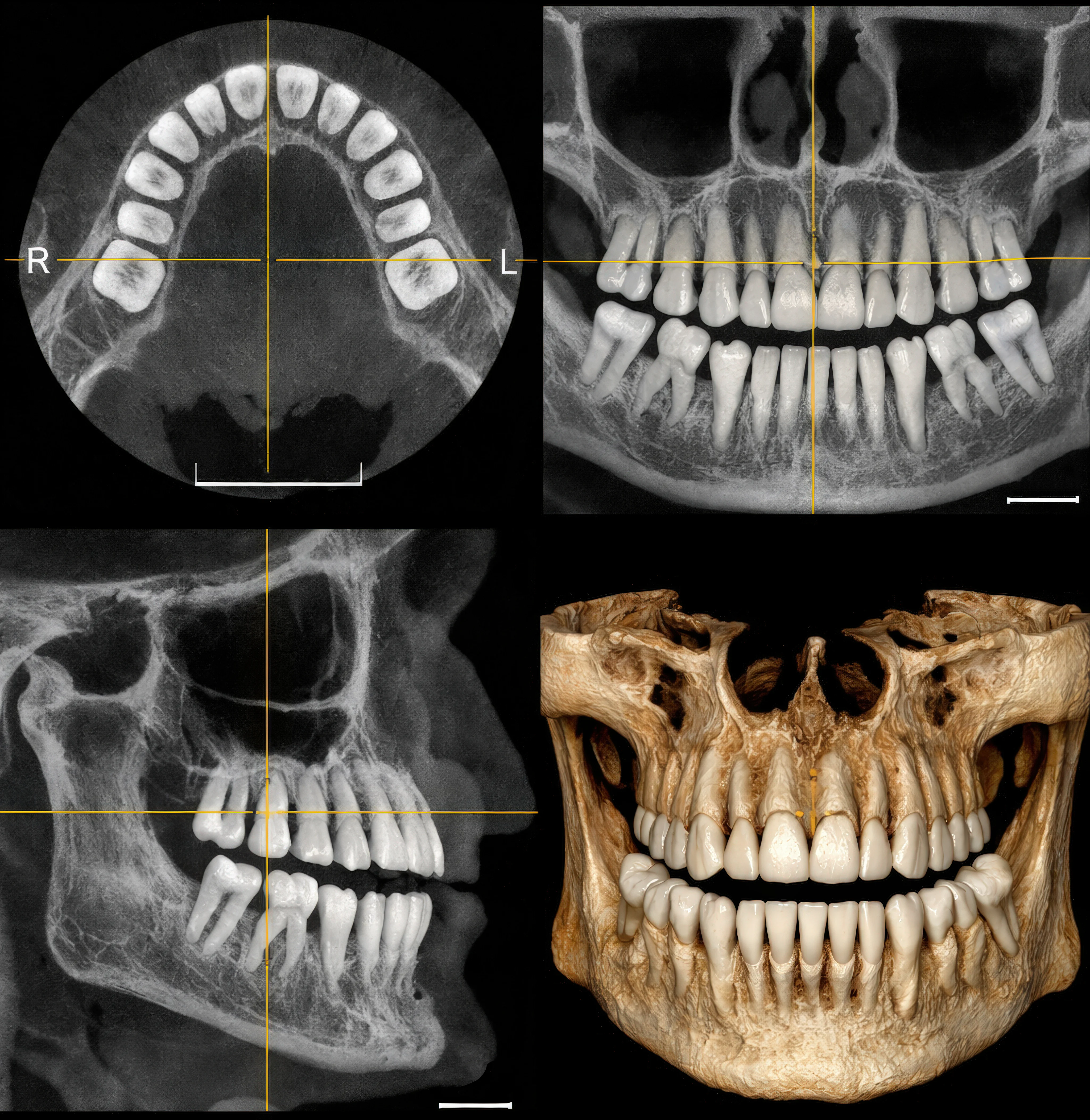
Cone Beam Imaging in Chicago, IL: A Patient Guide
Cone beam imaging at Armitage Oral Surgery in Chicago, IL provides a detailed 3D view of your teeth, jaws, and facial structures to support safe, accurate treatment planning. This advanced X-ray technology, often called cone beam computed tomography (CBCT), helps guide implants, wisdom tooth removal, jaw surgery, and more.
Cone Beam Imaging Explained
CBCT is a specialized dental scan that uses a cone-shaped X-ray beam to capture hundreds of images in seconds. Software then assembles those images into a 3D model of your mouth and jaw. Compared with a traditional 2D X-ray, the 3D view reveals the exact position of nerves, roots, bone, sinuses, and other anatomy that can be hidden in flat images.
Why Consider Cone Beam Imaging?
- Improved diagnostic clarity for fractures, root anatomy, cysts, and impacted teeth.
- Accurate implant planning with measurements of bone height, width, and density.
- Safer surgery by mapping the exact location of nerves and sinuses.
- TMJ and airway evaluation when 3D detail is helpful for diagnosis.
- Lower radiation dose than medical CT while delivering high-resolution detail for dentistry.
- Fewer surprises during procedures thanks to comprehensive planning.
How Cone Beam Imaging Works
- Preparation: You will remove glasses, earrings, dentures, and metal-containing accessories.
- Positioning: You are guided into place and asked to stay still for a few seconds.
- Scan: The machine rotates once around your head, capturing images in seconds.
- Results: Images are processed immediately and available for review the same day.
Have questions about 3D cone beam imaging in Chicago, IL, or need to schedule a CBCT consultation? Contact Armitage Oral Surgery at 773-486-2220 to schedule with Dr. Firas F. Katabi.
Ready for Advanced 3D Imaging?
Schedule your cone beam imaging consultation with Dr. Katabi in Chicago, IL. Get the detailed 3D view needed for precise diagnosis and treatment planning.
773-486-2220Frequently Asked Questions
A panoramic X-ray is 2D and best for a broad overview. A medical CT images large body areas with higher radiation. CBCT provides focused 3D detail of teeth and jaws for precise measurements and planning.
No. The scan is painless and quick, and the unit is open, not a tunnel. Most patients feel comfortable throughout.
No special fasting is needed. Remove metal items, wear comfortable clothing, and notify the team if you might be pregnant.
The scan takes seconds, and the full visit is often completed in minutes. Images are processed right away, so results are typically available the same day.
CBCT shows bone dimensions, nerve position, and anatomy in 3D. This helps select implant size and position, and it reveals fractures that 2D images can miss.
It can reveal the exact location of nerves, the thickness and height of bone, root fractures, hidden infections, and the relationship of impacted teeth to nearby structures.
The 3D model allows precise measurements, selection of implant size, and planning of the angulation and position to match your bone and final restoration.